MIT model E(3)-Pose tracks fetal head motion to sharpen prenatal brain MRI
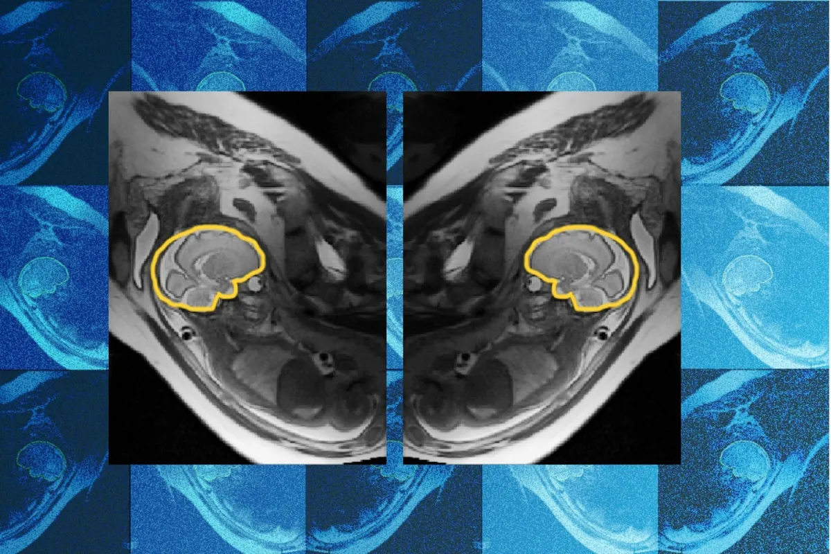
Researchers at MIT, Boston Children's Hospital and Inria have built a deep learning model called E(3)-Pose that tracks a fetus's head during MRI, aiming to produce complete, high-resolution brain images even when the baby moves. The model builds left-right symmetry directly into its architecture.
- E(3)-Pose is a deep learning model from MIT, Boston Children's Hospital and Inria that tracks fetal head motion during MRI.
- Left-right symmetry of the fetal head is embedded in the model architecture to help identify brain orientation.
- The paper was presented at the 2026 European Conference on Computer Vision.
- The model is mainly trained on third trimester fetuses; the team wants to extend it to the second trimester.
- Researchers say it could also apply to other hard-to-image anatomy and autonomous driving.
Why fetal movement ruins scans
A routine prenatal ultrasound can lead a sonographer to flag possible brain abnormalities for follow-up with magnetic resonance imaging (MRI). But the source notes that imperfect imaging can result in missed or incorrect diagnoses.
A fetal MRI is made of a series of 2D images, called slices, which stacked together form a high-resolution 3D image of the fetus's head. If the head shifts during the scan, each slice may be taken from a different angle. The result can be gaps in the final scan and unusual slice orientation. "One of the problems with having the baby move is that the slices are all over the place," says first author Ramya Muthukrishnan, an MIT PhD student in the lab of Polina Golland.
Those gaps matter clinically. "If you have coverage gaps, you can't see critical regions, and you need to be able to see well to detect abnormal development," Muthukrishnan explains.
Physics built into the network
E(3)-Pose helps image the fetal head while it is in motion so that a radiologist can examine a high-resolution result. What sets it apart, according to the researchers, is its focus on physics. Instead of training the model to learn physics from data alone, the principles are baked into the neural network. Left-right symmetry of the fetal head is embedded in the model architecture, which makes it easier for the model to identify the brain's orientation accurately.
The work was presented in a paper titled "Equivariant symmetry-aware head pose estimation for fetal MRI" at the 2026 European Conference on Computer Vision.
Muthukrishnan came to the project with a computer science background and no knowledge of MRI physics. Running the system on a real scanner proved difficult. "I was not prepared for how many times I would crash the scanner because putting things on hardware and getting it to work on hardware is a different beast than training a model to run on data on a GPU," she recalls.
She says her motivation came from shadowing a radiologist at Boston Children's Hospital. She saw a pregnant mother who felt uncomfortable after an hour in the MRI and quit the scan halfway, while the radiologist could not obtain a full, high-quality image.
The team and the clinical goal
Co-author Borjan Gagoski, an assistant professor of radiology at Harvard Medical School and a researcher at Boston Children's Hospital, developed a sequence that acquires additional MRI scans to help train the model. He also helped troubleshoot the scanner when it crashed. Co-author Aryn Lee recruited the pregnant volunteers and assisted with the scans. Senior authors include Elfar Adalsteinsson, Ellen Grant and Golland. Benjamin Billot of Inria, a former postdoc in Golland's group, helped Muthukrishnan get started with an initial method he developed.
Golland says the work "elegantly combines geometric concepts like equivariance and symmetry with the power of deep learning to change how MRI scanners acquire the images." Grant, who directs the Fetal Neonatal Neuroimaging and Developmental Science Center at Boston Children's, says it "will automate fetal brain MRI, replacing today's slow, manual process with faster and more comfortable scans for mothers, while also providing higher-quality images for radiologists to interpret."
Limits and what comes next
The model is primarily trained on third trimester fetuses. Muthukrishnan hopes to extend it to second-trimester or younger fetuses, which are "so small, they move around a lot more because there's more space in the uterus." She argues they matter more: "the earlier you detect brain abnormalities, the easier it is to intervene."
The researchers say E(3)-Pose could also help with imaging other hard-to-image anatomy, and even with autonomous driving, where limited sensors must capture noisy 3D data to track 3D objects.
For families and clinicians, the stated aim is fewer interrupted scans and clearer images for earlier diagnosis. The source does not report quantitative results or a timeline for clinical use. The work is supported by the MIT Jameel Clinic, the MIT Takeda Program, the Chou Family Transformative Research Fund, the National Institute of Biomedical Imaging and Bioengineering, and the Eunice Kennedy Shriver National Institute of Child Health and Human Development.