EPFL releases spora, the largest open atlas of spatial proteomics images
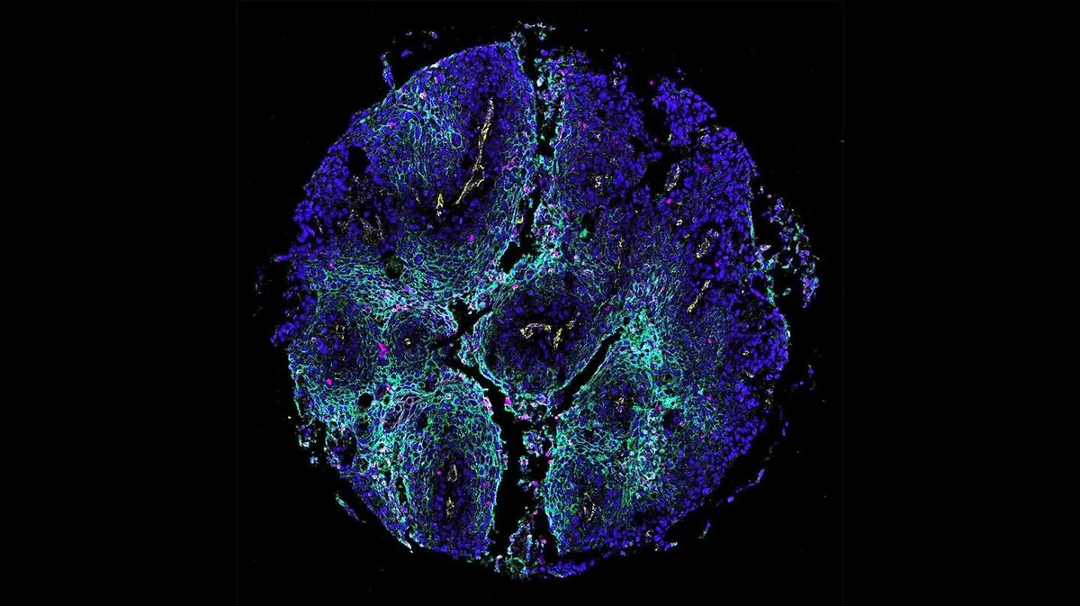
Researchers at EPFL have created spora, which they describe as the world's largest open-access collection of spatial proteomics tissue images, drawn from more than 5,000 patients. The resource is meant to speed up AI research into how tissues function and how diseases develop.
- EPFL released spora, which it calls the world's largest open-access spatial proteomics tissue image collection.
- It holds 13,546 multiplex images from 5,477 patients, across 14 tissue origins and five imaging technologies.
- Spora includes tools to access data, build AI models and test them across different studies.
- The project was built over two years with EMBL-EBI and Swiss hospitals and universities, as part of NAIPO.
- The team plans to add more tissues, diseases and imaging technologies.
Why tissue layout matters
According to EPFL, the arrangement of cells within tissue can reveal as much about disease as the cells themselves. When doctors examine a tumor, they want to know not only which cells are present but also how they are arranged and how they interact. The way cancer cells interact with neighboring immune cells can offer clues about how a disease develops or responds to treatment.
Spatial proteomics captures this organization by measuring dozens of proteins while preserving their locations. Because proteins help control what cells do, the images give a picture of how healthy and diseased tissues work.
The problem is that the resulting datasets are hard to combine. Laboratories around the world use different equipment, examine different proteins and store results in different ways. Merging them is complicated and time-consuming, which makes it difficult to use large amounts of data in conventional studies and to train AI models.
What spora contains
A team led by Charlotte Bunne's Artificial Intelligence in Molecular Medicine group at EPFL developed spora as a spatial proteomics reference atlas. It was released in collaboration with the European Molecular Biology Laboratory's European Bioinformatics Institute (EMBL-EBI).
- 13,546 multiplex images from 5,477 patients
- 14 tissue origins
- Five imaging technologies
- Paired histopathology images where available, linking molecular measurements with the tissue images used in routine pathology
Building it took two years of work by scientists and biomedical experts from EPFL, University Hospital Zurich, the University of Zurich, Geneva University Hospitals and the University of Geneva. They had to organize thousands of images, standardize protein annotations, match medical information and identify individual cells so that data from different studies could be compared.
"Gathering and harmonizing data across cohorts takes enormous time, creating a major hurdle for both large-scale AI development and biological discovery," says Benedikt von Querfurth, a doctoral student at EPFL and first author of the study. "We want to allow researchers to start with ready to use data so that they can focus on the models and biological questions."
Tools and testing across studies
The collection keeps links to the original studies and their annotations. Differences in the proteins measured, imaging conditions and available clinical information remain visible, which helps researchers decide which datasets suit their questions.
Beyond the atlas itself, spora offers tools to access and analyze the data, develop AI models and evaluate how well they perform across different studies. Lukas Klein, a postdoc at EPFL and the University Hospital Zurich, explains the reasoning: "This is important because a model can perform well within one study and still struggle when the patients, laboratory, or measurement technology change." He adds that shared datasets and evaluation protocols make it possible to examine those differences systematically.
Spora was developed as part of the National AI Initiative for Precision Oncology (NAIPO). It comes with AI models trained using large-scale computing resources through the Swiss AI Initiative. The work is described in a paper titled "spora: A Unified Multimodal Dataset and Platform for Spatial Proteomics", listed for NeurIPS 2026.
What it means for researchers and patients
For research groups, the main promise is time saved: they can start from harmonized, ready to use data rather than spending months aligning datasets. Spora currently focuses mainly on cancer research, but the team hopes to add more tissues, diseases and imaging technologies.
The longer-term aim is clinical. "Our goal is to turn what we learn across thousands of tissue samples into insights that can help individual patients," says Bunne. She says the team aims, working with clinicians, to translate these insights into better predictions of how a patient's disease will progress and which treatments are most likely to benefit them.
EPFL News: A new tissue atlas will help AI understand cancer